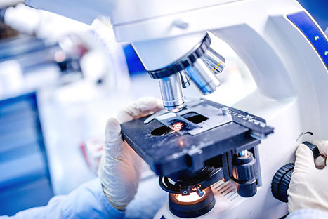
Materials Characterization
Facilities for characterization of energy-related materials:
Transmission Electron Microscope (TEM), JEOL JEM 1400
-
 Resolution (Lattice Image/ Point Image): 0.20 nm/ 0.38 nm
 -
Optimized take-off angle for best peak-to-background ratios and light element detection
-
UPRIGHT CONFOCAL MICROSCOPE, Leica TCS SP8 X
-
Upright Leica DM 6000 with adaptive focus, motorized XY-Stage (15 nm step size) and Super Z Galvo (1500µm/ 3 nm step size)
-
Tandem scanner 8 KHz:
-
Detection range: 400-800 nm
-
Internal detection channels: 2xPMT, 2x HyD
-
Transmitted light detectors: BF (Brightfield) prism for DIC measurement in confocal mode
-
Equipped with Tokai Hit Stage Incubator providing 37˚C and 5% CO2 for live cell imaging
Scanning Probe Microscope with Hysitron Attachment, Bruker Dimension ICON
-
Materials mapping
-
Nanomechanics characterization
-
Nanoelectrical characterization
-
Biological characterization
AMG EVOS FL Microscopy
-
Light cubes: DAPI (Ex 360nm/ Em 447nm), GFP (ex 470 nm/ Em 525 nm), RFP (Ex 530nm/ Em 593 nm), White (for non-transparent samples)
-
Objectives: 4x, 10x, 20x, 40x LWD objectives and 100x coverslip-corrected oil objective
-
Equipped with Bioptechs stage temperature controller providing 37˚C for live cell observation
-
THERMOMECHANICAL CHARACTERIZATION
-
Thermal Gravimetric Analysis (TGA, TA Q50):
-
Temp. range: ambient+5~1000oC
-
Differential Scanning Calorimetry (DSC, TA Q2000):
-
Temp. range: -90~550 oC
-
Dynamic Mechanical Analysis (DMA, TA Q800): Temp. range: -145~600 oC
-
Thermal Conductivity Meter (DTC-300): Temp. range: -20~300 oC;
-
Thermal conductivity range: 0.1~40 W/m.K
Spectroscopic Ellipsometry, Horiba UVISEL FUV
-
Thin film thickness from 1 Å to 30 µm
-
Surface and interface roughness
-
Optical constants (n, k) for isotropic, anisotropic and graded films
-
Characterization of thickness and optical constants in the VIS-FUV spectral range of thin films and multilayer stacks for:
-
Dielectrics
-
High k, low k materials
-
Photo resists
-
Plastics
-
Amorphous semiconductors
-
Thin metal films
-
Glass
-
Polymers
TEM Sample Preparation
-
Leica EM UC7 with Cryo Attachment
-
High quality ultra-microtome for precise room temperature and cryo sectioning (-15~ -185˚C)
Thermo Scientific Nicolet iS50 FTIR Spectrometer
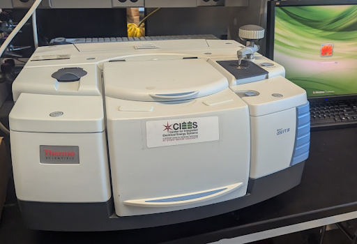 The Nicolet iS50 FTIR Spectrometer is dual source-capable, offering users both the
Polaris long-life IR source and a tungsten-halogen white light source. Full support
for a wide range of accessories offers the flexibility needed to target the spectral
analysis.
The Nicolet iS50 FTIR Spectrometer is dual source-capable, offering users both the
Polaris long-life IR source and a tungsten-halogen white light source. Full support
for a wide range of accessories offers the flexibility needed to target the spectral
analysis.
Colloidal Dynamics ZetaProbe Analyzer
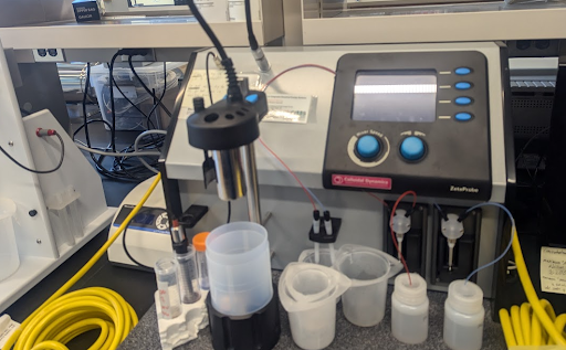 The ZetaProbe features a compact design with built-in titration, versatile dip probe
sensor, and software wizards. Automatic titration offers unattended rapid isoelectric
point determination, as well as optimum dispersant or flocculant level control in
a click of a button. The ZetaProbe offers many advances not found in other analyzers
including automatic correction for particle size and double layer distortion.
The ZetaProbe features a compact design with built-in titration, versatile dip probe
sensor, and software wizards. Automatic titration offers unattended rapid isoelectric
point determination, as well as optimum dispersant or flocculant level control in
a click of a button. The ZetaProbe offers many advances not found in other analyzers
including automatic correction for particle size and double layer distortion.
Quantachrome – BET Surface Area and Pore Size Analyzer
 Brunauer-Emmett-Teller (BET) surface area analysis is the multi-point measurement
of an analyte’s specific surface area (m2/g) through gas adsorption analysis, where
an inert gas such as nitrogen is continuously flowed over a solid sample, or the solid
sample is suspended in a defined gaseous volume. Small gas molecules adsorb to the
solid substrate and its porous structures due to weak van der Waals forces, forming
a monolayer of adsorbed gas. This monomolecular layer, and the rate of adsorption,
can be used to calculate the specific surface area of a solid sample and its porous
geometry.
Brunauer-Emmett-Teller (BET) surface area analysis is the multi-point measurement
of an analyte’s specific surface area (m2/g) through gas adsorption analysis, where
an inert gas such as nitrogen is continuously flowed over a solid sample, or the solid
sample is suspended in a defined gaseous volume. Small gas molecules adsorb to the
solid substrate and its porous structures due to weak van der Waals forces, forming
a monolayer of adsorbed gas. This monomolecular layer, and the rate of adsorption,
can be used to calculate the specific surface area of a solid sample and its porous
geometry.